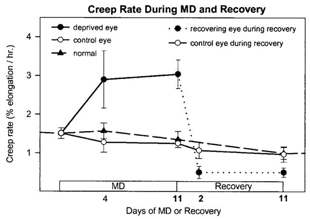
Figure 1.

Summary of scleral extensibility (creep rate) during monocular deprivation (MD) and during recovery from MD (replotted from Siegwart and Norton20). Creep rate was increased in the deprived eyes, compared with control and normal eyes, after 4 days of MD and remained high after 11 days of MD. Two days after recovery was initiated by removing the diffuser, the creep rate in the deprived eyes (dashed line) was significantly lower than in the control eyes. Creep rate was measured in 3-mm wide strips of sclera under 1 gram of constant tension, which approximates the tension produced by normal intraocular pressure (n = 3 animals, each group). In normal animals, left and right eyes were averaged.
