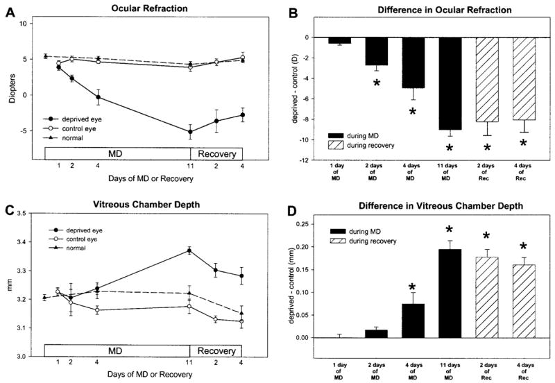
Figure 2.

Ocular refraction and vitreous chamber depth during MD and recovery. Compared with their fellow control eyes, the deprived eyes became increasingly myopic (A) and their vitreous chambers became increasingly deep (C) as a function of days of MD. During recovery, the progression of myopia and the increase in vitreous chamber depth abruptly reversed. Data from normal animals (average of left and right eyes) are shown for comparison. (B, D) The difference between the deprived and control eyes, respectively (n = 5 in each group). Results are expressed as the mean ± SEM (*P < 0.05).
