
Figure 4.
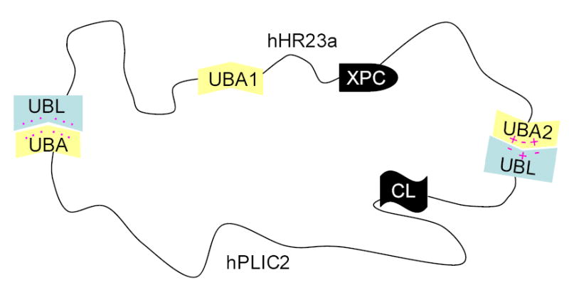
A schematic model of how hHR23a interacts with hPLIC2. A hydrophobic surface of hHR23a’s UBL domain interacts with hPLIC2’s UBA domain, whereas charged residues appear to play a role in its C-terminal UBA domain binding to hPLIC2’s UBL domain. Pink dots represent hydrophobic surfaces, whereas the “+” and “−“ symbols reflect electrostatic surfaces.