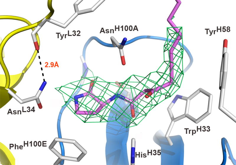
Figure 2.

Antibody combining site of RS2-1G9 bound to an AHL lactam mimetic (pink). The light and heavy chains are colored in yellow and blue, respectively. The σa-weighted 2Fo-Fc electron density map around the ligand is contoured at 1.4σ. The microenvironment at the very bottom of the binding pocket tolerates high-affinity binding of both the lactam and the lactone, since AsnL34 does not form a hydrogen bond to the NH-group of the lactam. A potential hydrogen bond in the lactone complex model with the NH2-group of AsnL34 may account for its increased affinity with respect to the lactam. The displayed orientation of the terminal amide group of AsnL34 is preferred due to formation of a hydrogen bond with TyrL32. CDR L3 is omitted for clarity.
