
Figure 3.
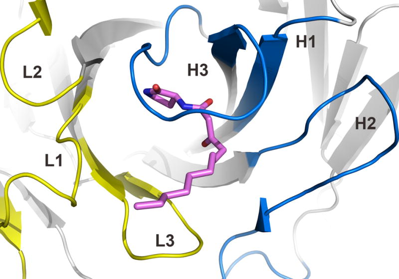
Architecture of the antibody combining site of RS2-1G9. The CDRs of light and heavy chains are highlighted in yellow and blue, respectively. The lactam 3 is shown in pink. The tip of the CDR H3 loop bends over the ligand and largely seals it from bulk solvent.