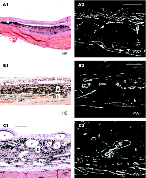
Figure 1 (A1–C1) Haematoxylin and eosin (HE) staining with accompanying von Willebrand factor (VWF) staining (A2–C2) of three retinal pigment epithelium–choroid grafts: vertically bridging vessels between recipient and graft. Arrows indicate bridging vessels; g, graft; f, fibrovascular layer; h, host choroid; t, transition healthy‐degenerated retina; v, intraretinal or intrachoroidal vacuoles filled with silicone oil. Scale bars: A1–C1, 200 μm and A2–C2, 100 μm.

An official website of the United States government
Here's how you know
Official websites use .gov
A
.gov website belongs to an official
government organization in the United States.
Secure .gov websites use HTTPS
A lock (
) or https:// means you've safely
connected to the .gov website. Share sensitive
information only on official, secure websites.
