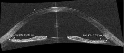
Figure 3 Anterior chamber image with SL‐OCT. Direct angle visualisation is possible; software provides automated quantification of angle parameters. The values for the angle opening distance at 500 μm (AOD 500) and the trabecular‐iris spur area at 500 μm (TISA 500) can be seen. The asterisk (*) shows the presence of a contact lens.

An official website of the United States government
Here's how you know
Official websites use .gov
A
.gov website belongs to an official
government organization in the United States.
Secure .gov websites use HTTPS
A lock (
) or https:// means you've safely
connected to the .gov website. Share sensitive
information only on official, secure websites.
