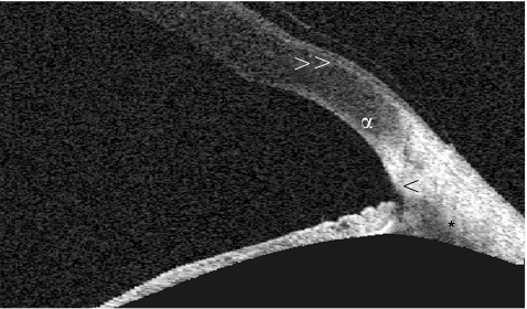
Figure 5 High‐resolution scan of the anterior chamber angle with the Visante OCT demonstrating angle visualisation. The arrowhead (<) shows the scleral spur and the asterisk (*) the anterior ciliary body. The letter α marks the limbal transition from cornea to sclera and the double arrowhead (>>) points to Bowman's membrane of the cornea with the overlying epithelium.

An official website of the United States government
Here's how you know
Official websites use .gov
A
.gov website belongs to an official
government organization in the United States.
Secure .gov websites use HTTPS
A lock (
) or https:// means you've safely
connected to the .gov website. Share sensitive
information only on official, secure websites.
