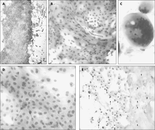
Figure 1 Histological and immunohistochemical assessment of whole‐mounted contact lenses. Contact lenses (lotrafilcon A, A–E) were removed from patients (A and B, patient 1; C, patient 2; D, patient 3; and E, patient 4) who had undergone surgery for pterygium resection. Contact lenses were flat mounted and assessed for keratin 15 expression (A and B), presence of mucin (C, periodic acid‐Schiff), morphological appearance (D, haematoxylin and eosin), and proliferative capacity (E, p63). Immunoreactivity is denoted by the red cytoplasmic (A and B) or nuclear (E) staining. Some contact lenses were counterstained with haematoxylin (A and B) while others were not (E) to avoid masking any nuclear immunoreactivity. The asterisks (*) in panel A indicate the contact lens serial number, which is visible but out of focus. The arrows in panel E point to intensely stained, whereas the arrowheads identify moderately stained p63‐positive cell nuclei. Original magnification ×40 (A), ×200 (B and E), ×400 (C and D).

An official website of the United States government
Here's how you know
Official websites use .gov
A
.gov website belongs to an official
government organization in the United States.
Secure .gov websites use HTTPS
A lock (
) or https:// means you've safely
connected to the .gov website. Share sensitive
information only on official, secure websites.
