
Figure 1.
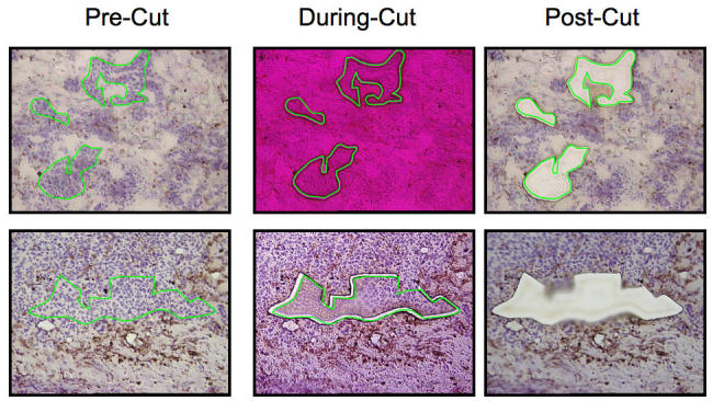
Frozen sections of OC were prepared and stained with H&E. The lines indicate the areas selected for microdissection, then this group of cancer cells is microdissected using a low power pulse laser. The figure shows two different samples before (pre-cut), during (during-cut), and after (post-cut) microdissection.