
Figure 2.
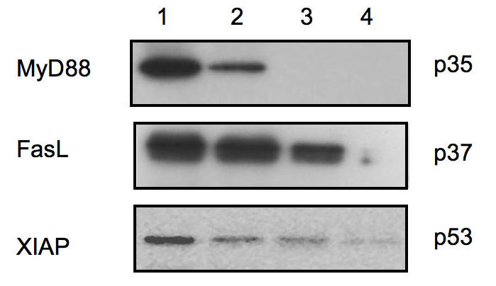
Western blot analysis for microdissected ovarian cancer cells. Eight microns thick frozen sections of epithelial ovarian tumors were prepared and stained with H&E. Different numbers of pure cancer cells were microdissected and evaluated for the expression of FasL, XIAP, and MyD88. 1 = 8,000 cells; 2 = 5,000 cells; 3 = 1000 cells; 4 = 100 cells. As shown, a strong signal was observed for MyD88 expression in samples containing 8,000 and 5, 000 microdissected cells, and no signal was detected for samples containing less than 1000 cells. For FasL and XIAP, a signal can be observed with as little as 1000 cells.