
Figure 5.
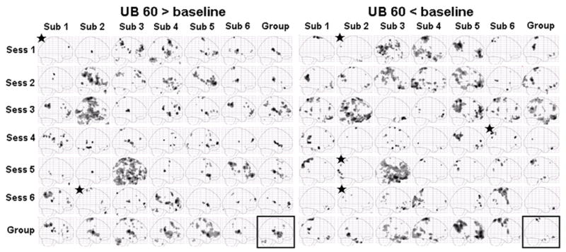
Sagittal maximum intensity projections (MIPs) for electroacupuncture stimulation at UB 60 in both individual session and group analysis. The subject average for all 36 sessions is shown in the box at the bottom right. The group averages across all six sessions for each subject are shown along the bottom row. The group averages across the 6 subjects in each session are shown in the far right column. Signal increases are shown on the left and signal decreases on the right. Star indicates no activation was observed at the original threshold of p<0.0001 uncorrected with 10 contiguous voxels, a lowered threshold of p<0.001 uncorrected with 10 contiguous voxels was used in this case.