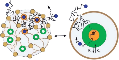
Fig. 2.
Homogenization of the PSD. (Right) Scattered free, bound, and scaffolding molecules and other obstacles and fences in the PSD. (Left) A course-grained model with concentrated free (green), bound (orange), and scaffolding and other molecules (brown) (see text). [Reproduced with permission from ref. 29 (Copyright 2006, Biophysical Society).]

