
Fig. 25.
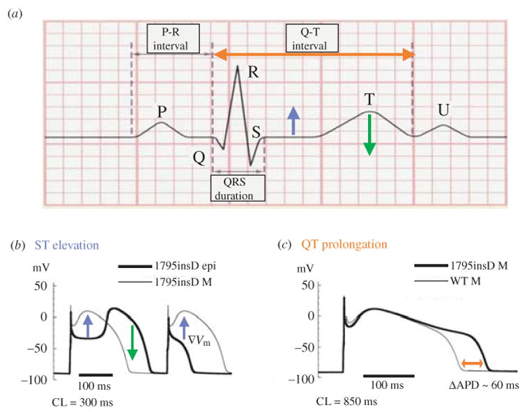
Mechanisms of ECG changes caused by the 1795insD mutation. (a) Stylized ECG defining the different deflections, waves and intervals. (b) Cellular mechanism of ST elevation and T-wave inversion. At fast rate, loss of dome or coved dome in epicardial cells creates a voltage gradient (▽Vm, blue arrows) between these cells (bold line) and M cells (thin line) during the AP plateau, leading to ST-segment elevation on the ECG [blue arrow in (a)]. Greatly prolonged notch in a coved-dome epicardial AP could delay its repolarization beyond that of M cell, thus reversing the normal gradient (normally, M cells have the longest APD and repolarize last) as indicated by the green arrow in (b), possibly leading to T-wave inversion [green arrow in (a)]. (c) Cellular mechanism of QT prolongation. At slow rate M-cell APD is prolonged by the mutation (red arrow). The delayed repolarization is reflected as QT prolongation on the ECG [red arrow in (a)]. [Simulation data in (b) and (c) are reproduced from Clancy & Rudy, 2002, with permission.]