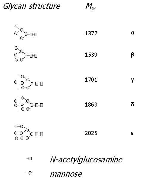
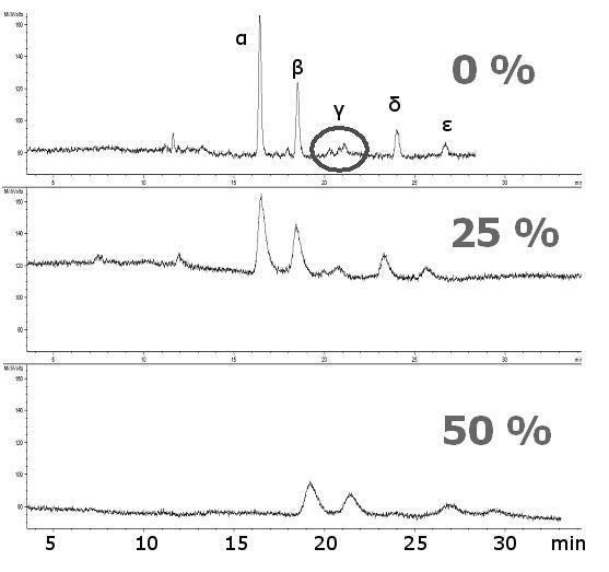
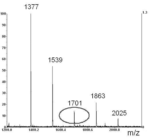
Figure 4.


Effect of the amount of copolymerization ligand (x %) in a polymerization feed on separations of glycans cleaved from ribonuclease B. Structures of glycans and their corresponding MALDI-TOF-MS. Sample: Ribonuclease B glycans tagged with 2-AB, ca. 10 μg/20 μl, inj. 8 kV/10 s; Mobile phase: ACN/ 240 mM NH +4 formate/H2O (60:1:39), CEC-LIF, ∼ 600 V/cm, ∼ 3.5 μA; Columns AAm/Bis (T 5%, C 60%, TRIS-AAm x%), (ltot = 40.0 cm, ldet = 33.0 cm); (A) 0 % (blank monolith), (B) 25 % TRIS-AAm, (C) 50 % TRIS-AAm
