
FIGURE 1. STRUCTURE OF HUMAN RHINOVIRUS 14.
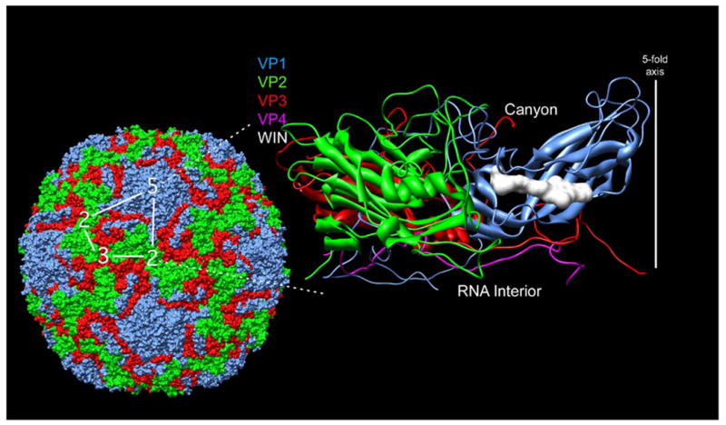
Shown here is the structure of the entire human rhinovirus 14 capsid5 (left) and of the protomeric unit (right). The capsid proteins VP1-4 are colored blue, green, red, and mauve, respectively. The protomeric unit, is outlined in white on the whole particle at the left. The image on the right is a ribbon representation of the protomeric unit shown with the outer surface towards the top and the RNA interior towards the bottom. The antiviral, WIN, compound is shown here as a white space filling model as it is bound in a cavity beneath the canyon floor3. The program used for the visualization was Chimera57.