
FIGURE 3. FAR-UV CIRCULAR DICHROISM OF HRV14 UNDER UREA TREATMENT.
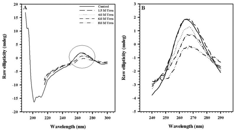
Changes in secondary structure of HRV14 treated with urea. In A, the entire far-UV spectra are shown; in B, the RNA region of the spectra is enlarged. The virus concentration was 50 μg/mL. The spectra were obtained in 50 mM bis-Tris propane buffer pH 7.5, using a 0.1-cm path length quartz cell.