
FIGURE 5. MALDI TOF-MS ANALYSES AND TRYPSIN DIGESTION OF HRV14 WITH AND WITHOUT DRUG.
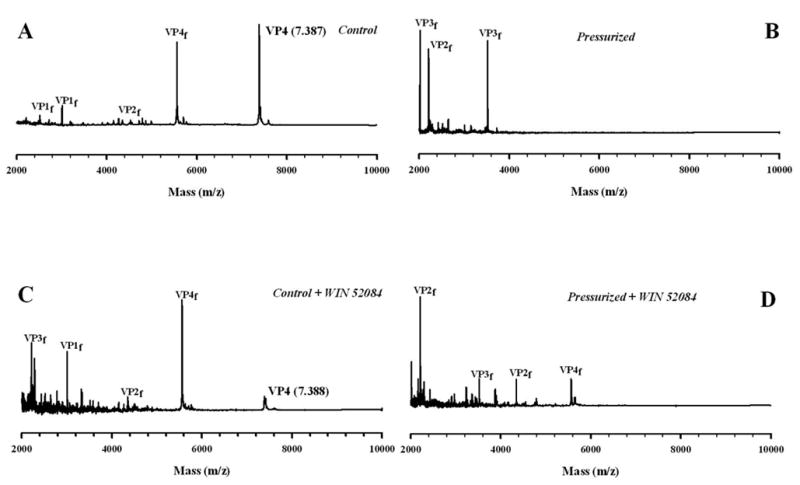
Trypsin digestion time course (10 minutes) of HRV14 after overnight pressurization at 3.2 kbar. In A, HRV14 was incubated with trypsin for 10 min at 25°C. In B, the virus was incubated with trypsin after overnight pressurization at 3.2 kbar. In C, the virus was digested in the presence of WIN 52084 (10 μg/mL) and D, the virus with drug was digested after pressurization overnight at 3.2 kbar. The fragments of proteins VP1, VP2, VP3 and VP4 are shown as f. All spectra are displayed on the same intensity scale. Drug and virus were present at a final concentration of 10 μg/mL and 1 mg/mL, respectively. The virus was diluted in 10 mM Tris buffer, pH 7.6. The spectra were recorded at 25 °C. In each case the digestion mixture was filtered using a Centricon system.