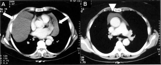
Fig. 1 Axial contrast-enhanced computed tomographic scans of the chest (mediastinal window) at the level of the left ventricle (A) and the aorticopulmonary window (B) show a non-enhanced, low-attenuated, well-circumscribed, dumbbell-shaped mass adjacent to the heart (arrows). Two components of the cyst are joined to each other in front of the ascending aorta (arrowhead) at the aorticopulmonary level.
