
Fig. 3.
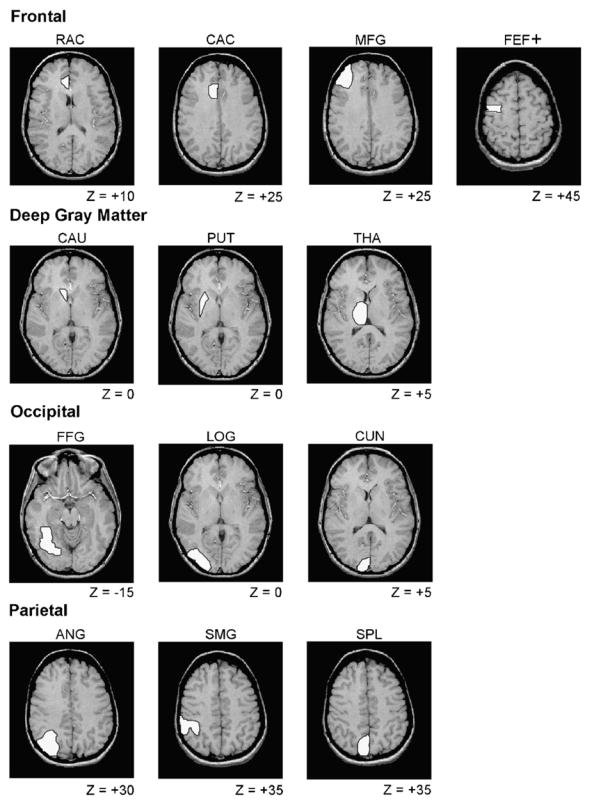
Examples of gray matter ROIs. For each ROI, an example is displayed of one representative T1-weighted MR slice, with Z value (in mm) for location of this slice relative to the AC–PC plane. Only the left hemisphere ROIs are illustrated, but all ROIs were drawn in both hemispheres. Although only one slice is illustrated, each ROI could include multiple slices (see Table 1), and the ROIs were defined for each participant individually. RAC, rostral portion of anterior cingulate; CAC, caudal portion of anterior cingulate; FEF+, frontal eye field; MFG, middle frontal gyrus; CAU, caudate; PUT, putamen; THA, thalamus; CUN, cuneus; FFG, fusiform gyrus; LOG, lateral occipital gyri; ANG, angular gyrus; SMG, supramarginal gyrus; SPL, superior parietal lobule. We use the label FEF+ to distinguish our ROI in BA 6 from the frontal eye field defined classically in BA 8 [29].