
Figure 1.
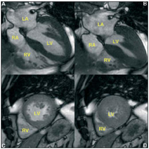
Cardiac MRI images in a subject with increased LV mass. A-B. Four-chamber view demonstrating the four cardiac chambers, in end-diastole (A) and end-systole (B). C-D. Short-axis view demonstrating the ventricles, in end-diastole (C) and end-systole (D). LA=left atrium, RA=right atrium, LV=left ventricle, RV=right ventricle.