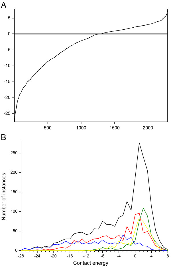
Figure 5.
Distribution of contact energies for mutated amino acids. The energies were calculated with the RankViaContact program. A) Data for all mutations, and B) distribution in secondary structural elements α-helices (red), β-strands (blue), turns and bends (green), outside secondary structures (yellow), and whole proteins (black).

