
FIG. 9.
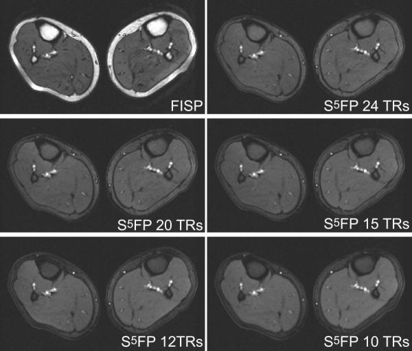
Comparison of im'ages of calf acquired using conventional FISP and S5FP pulse sequences with various train-lengths. The S5FP images employed opening and closing subsequences of 5 pulses and 1 pulse, respectively, a water-fat separation angle of 90°, and RF spoiling between successive SSFP trains.