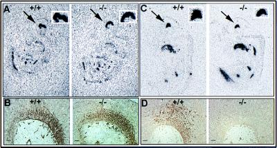
Figure 4.
Analysis of the phenotype of cells in the ventral midbrain of wild-type and Nurr1 mutant embryos. (A) Autoradiographic localization of hybridization to the ventral marker, HNF3β. The arrow shows the strong staining in the ventral part of the midbrain of both 12.5 days wild-type (Left) and mutant (Right) mice embryo. The square on the top shows a higher magnification of this expression. (B) Immunostaining for the general neuronal marker, 3A10, in 11.5 day wild-type and mutant mouse embryos. (C) In situ hybridization analysis of Ptx-3 mRNA expression in 11.5 day mouse embryo. The arrow indicates the positive staining in the ventral midbrain. Wild-type and mutant mice showed similar staining. (D) Immunohistochemical localization of TH expression in the ventral midbrain of 12.5 day wild-type embryo and lack of expression in the nurr1−/− midbrain. (Bars = 20 μm.)

