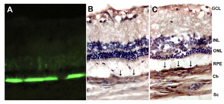
Figure 1. VEGF and GFP expression in rat eyes 8 weeks after AAV-VEGF or AAV-GFP injection.

Eyes were fixed, embedded in OCT medium, sectioned (10μm) and stained. A. GFP strongly expressed in RPE and faintly expressed in photoreceptors from an eye injected with AAV-GFP. B. In AAV-GFP injected eye, there is relatively little DAB staining in RPE (black arrows). C. In AAV-VEGF -injected eye, there is intense DAB staining at the RPE (black arrows), indicating VEGF overexpression. GCL, ganglion cell layer; INL, inner nuclear layer; ONL, outer nuclear layer; RPE, retinal pigment epithelium; Ch, choroid; Sc, sclera.
