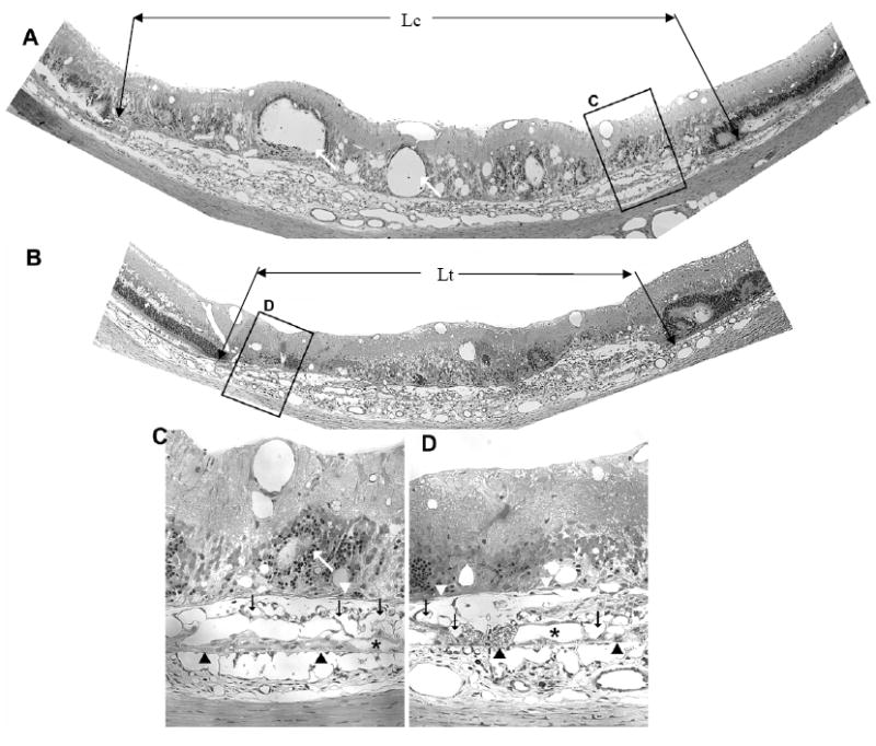
Figure 2. AG013764 reduces VEGF-induced CNV in SD rats.

Panels A and B are 1.5 μm sections from eyes injected intravitreally with CMC carrier (control) or AG013764 (three 20 μg injections at 5 day intervals). Compared with the control eye, sections from the AG013764 treated eye show less blood vessel proliferation in the SRS (black arrows), areas of disorganized photoreceptors (white arrows), RPE proliferation (white arrowheads), and RPE “lakes” (asterisks). Bruch’s membrane indicated by black arrowheads. Panels C and D are sections in A and B at higher magnification.
