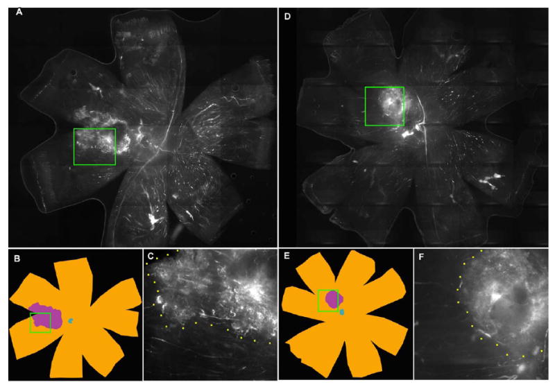
Figure 5. FITC-dextran whole mount of eyes intravitreally injected with AG013711 or control.

Animals injected every five days for two weeks starting at six weeks after AAV-VEGF injection. Panels A, B, C from a control eye injected with carrier and panels D, E, F from AG013711 injected eye. Panels A and D, photographs of whole eye flat mount. Panels B and E, typical sets of contours drawn using the Neurolucida system. The total flat mount area (≈ 50 mm2) is colored brown and the blue center denotes the optic nerve. Area of CNV shown in purple. Panels C and F, insets from A and D at higher magnification, show a typical boundary between areas of proliferation (CNV) and non-proliferation.
