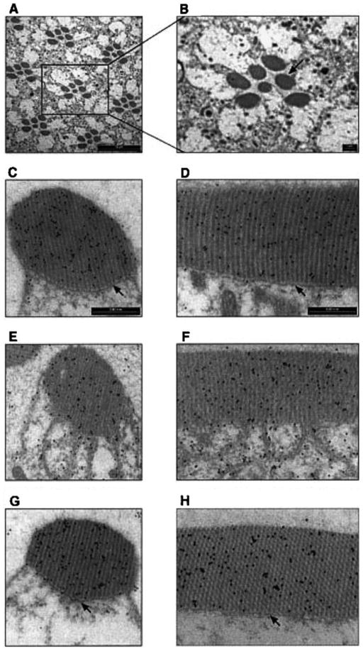
Fig. 1.

Light-dependent translocation of DGqα and rhabdomeral architectural changes shown by immunogold labeling. Panels A and B show a low magnification cross-section of the Drosophila’s compound eye for orientation. The seven dark oval structures of each visual unit (ommatidium) are the rhabdomeres, the signaling compartment of the photoreceptor cell composed of tightly packed microvilli. Panels C, E, G show cross-sections of a single rhabdomere while panels D, F, H show a longitudinal section along the axis of the rhabdomere. In these panels, DGqα is localized by immunogold labeling, seen as dark dots. Panels C and D are from dark-adapted flies. Panels E and F are from flies illuminated for 60 min with blue light. Panels G and H are from flies that had been illuminated for 60 min and subsequently kept for 2 h in the dark. Illumination (panels E and F) caused remarkable architectural changes in the cell. The boundary between the rhabdomere and the rest of the cell disappears due to disruption of the cytoskeletal Rhabdomeral Terminal Web (RTW—marked with arrows in panels C and D). As a result, the rhabdomere collapses and membrane tendrils resembling a house painter’s brush are seen intruding into the cytosol. These architectural changes are completely reversible, as after 2 h in the dark (panels G and H) the rhabdomeres are indistinguishable from those of dark-adapted flies (panels C and D). Similar observations were found in six different flies. (From Kosloff et al., 2003.)
