
Fig. 2.
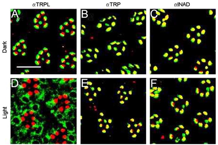
Light-dependent translocation of TRPL molecules in the photoreceptor cells of Drosophila compound eyes. Cross-sections through wild-type Drosophila (A–F) eyes of light- and dark-raised flies were double labeled with rhodamin-coupled wheat germ agglutinin, which specifically labels rhabdomeral photoreceptor membranes (red fluorescence), and antibodies against TRPL, TRP, and INAD, as indicated. The overlay of both markers appears yellow. Scale bars in (A), 10 μm. (Modified from Bähner et al., 2002.)