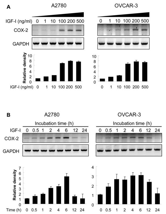
Fig. 1. IGF-I upregulates COX-2 mRNA expression.

(A) A2780 and OVCAR-3 cells were cultured in serum-free medium for 16 h, then treated with the indicated concentrations of IGF-I for 6 h. COX-2 and GAPDH mRNA levels in the cells were detected by RT-PCR. (B) The cells were treated with 200 ng/ml of IGF-I for 0 to 24 h as indicated. COX-2 and GAPDH mRNA expression was analyzed by RT-PCR. The levels of COX-2 mRNA were quantified using NIH Image J software, and normalized to those of GAPDH levels. The mean ± SD of the corresponding densitometry data from replicate experiments were showed in lower panels of each figure.
