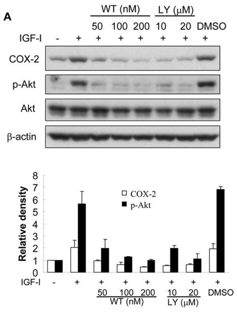
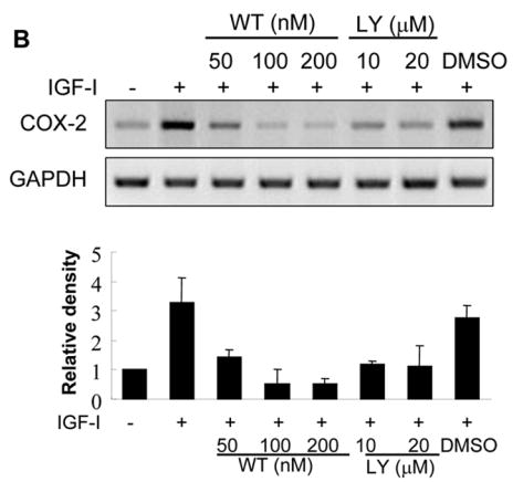
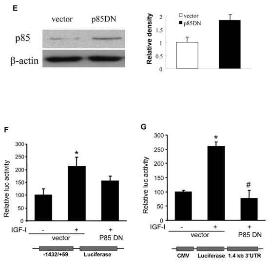
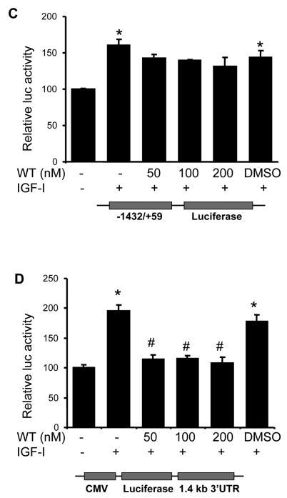
Fig. 4. PI3K signaling is required for IGF-I-induced COX-2 expression.


(A) Serum-starved A2780 cells were pretreated with wortmannin (WT), LY294002 (LY) or the solvent DMSO followed by the incubation with 200 ng/ml IGF-I for 8 h. COX-2, phospho-Akt (p-Akt, Ser473), total Akt, and β–actin protein levels were analyzed by immunoblotting. The COX-2 protein signals were normalized to those of β-actin levels, and p-Akt signals were normalized to those of total Akt levels. The mean ± SD of the corresponding densitometry data from duplicate experiments were shown in the lower panel. (B) The cells were pretreated with wortmannin (WT), LY294002 (LY) or solvent (DMSO) followed by incubation with 200 ng/ml IGF-I for 6 h. COX-2 and GAPDH mRNA levels were detected by RT-PCR. The COX-2 mRNA signals were normalized to those of GAPDH levels with the mean ± SD of the corresponding densitometry data from duplicate experiments shown in the lower panel. (C and D) The cells were transfected with phPHES2(−1432/+59) and Luc-3′UTR, respectively; and cultured overnight. The cells were switched to serum-free medium in the absence or presence of wortmannin or 200 ng/ml IGF-I for 16 h. Relative luciferase activity was the ratio of luciferase/β-gal activity, and normalized to that of the control cells. Data are presented as mean ± SD from three independent experiments. * indicates that the value is significantly different when compared to that of the control (p<0.05); # indicates that the value is significantly different when compared to that of IGF-I treatment alone (p<0.05). (E) A2780 cells were transfected with empty vector or a vector expressing dominant-negative PI3K construct (p85DN). After 24 h, the cells were collected and lysed in RIPA buffer. Total proteins were analyzed by immunoblotting using antibodies against p85 and β-actin. The right panel shows the relative densitometry data of p85 protein signals normalized to those of β-actin levels. (F) A2780 cells were co-transfected with phPHES2(−1432/+59), pCMV-βgal, empty vector, or a vector expressing dominant-negative PI3K construct (p85DN); and cultured overnight. The cells were switched to serum-free medium, and incubated in the absence or presence of 200 ng/ml IGF-I for16 h. (G) The cells were co-transfected with Luc-3′UTR, pCMV-βgal, empty vector, or a vector expressing p85DN. The cells were switched to serum-free medium and incubated in the absence or presence of 200 ng/ml IGF-I for 16 h. Relative luciferase activity was analyzed as described above. *indicates that the value is significantly different when compared to that of the control (p<0.05); # indicates that the value is significantly different when compared to that of the vector-transfected cells treated with IGF-I alone (p<0.05).

