
Figure 1.
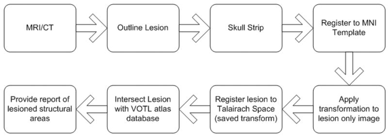
Block diagram depicting processing steps for lesion localization of a single subject’s MRI or CT scan. The lesion is manually outlined slice by slice. The brain is automatically skull stripped allowing for a more accurate spatial normalization. The brain is then automatically registered to the MNI Template brain. The transformation matrix is saved and applied to an image of the lesion only. If the AAL atlas option is chosen, the intersection of the lesion with the AAL atlas is determined and a report is produced. If the VOTL atlas option is chosen, another spatial transformation is applied, which maps the MNI template space to the Talairach space of Talairach/Tournoux 1988. The lesion image is compared to the VOTL atlas database and anatomic structures that intersect the lesion are reported.