
Figure 2.
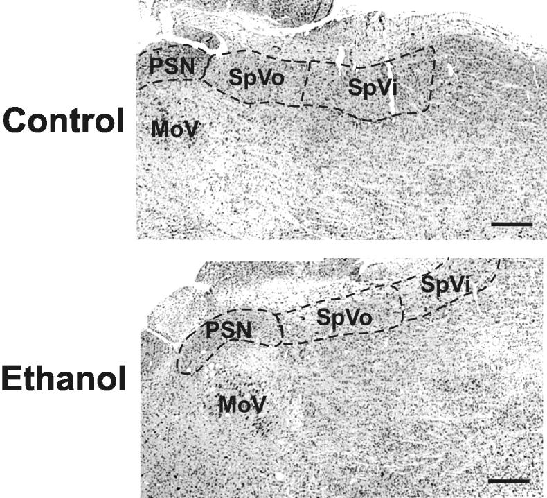
Appearance of the sensory cranial nerve nuclei
Three sensory trigeminal nuclei were identifiable in horizontal sections stained with cresyl violet. Samples representing the animals treated with saline (controls) or ethanol were taken from cohorts dosed on gestational day 8. No gross differences were apparent between the groups. Rostral is oriented to the left and lateral to the top. Scale bars are 500 μm.