
Figure 9. DTI-based Prediction of Normal Histology in the Anterior Commissure Following TBI.
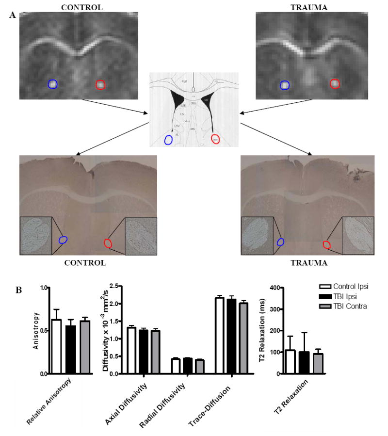
(A) DTI images showing RA in the anterior commissure. The APP stained regions show no positive staining that would indicate injury (4x; inset 10x). (B) There were no significant changes in any of the DTI or conventional MRI parameters in the anterior commissure following trauma.