
Fig. 1.
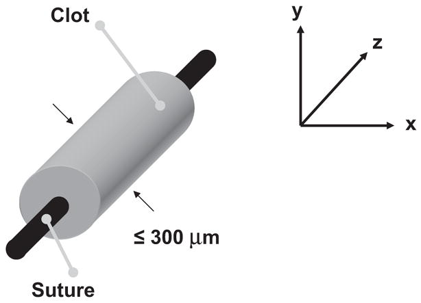
Typical sample blood clot orientation for this study. Using a 20 μl micropipette and a 7-0 suture, a blood clot with a volume of approximately 5–8 μl is created. The clot is approximately 300 μm in width, allowing it to be visualized completely within the field-of-view of the microscope using a 40× objective.