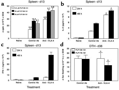
Figure 4.
Preclinical administration of anti–VLA-4 does not inhibit activation of peripheral PLP139-151–specific T-cell responses. Splenic lymphocytes from three mice per group were harvested day 13 after immunization and tested for PLP139-151–specific proliferative and Th1 cytokine responses. Three treatments with either anti–VLA-4 or control Ab had been administered by this time point. (a) Viable cells (5 × 10⁵/well) were cultured with the indicated concentrations of PLP139-151 for 4 days. Cultures were pulsed with 3H-TdR 20 hours before harvest. Data are presented as Δ cpm (3H-TdR incorporation in cultures containing peptide antigen, 3H-TdR incorporation in cultures containing medium). Stimulation indices (3H-TdR incorporation in cultures containing peptide antigen, 3H-TdR incorporation in cultures containing medium) are indicated above each bar. 3H-TdR incorporation in cultures containing medium only were 2,849 ± 183 and 8,850 ± 1,227 for control and anti–VLA-4 groups, respectively. IL-2 (b) and IFN-γ (c) levels in the supernatants of cultures harvested at 24 and 48 hours after stimulation with 25 μM of PLP139-151 were determined by ELISA as described in Methods. (d) DTH responses to both the initiating PLP139-151 peptide and the relapse-associated PLP178-191 peptide were evaluated in 4–5 mice treated with either control Ig or anti–VLA-4 at day 36 after immunization. Data represent the mean 24-hour change in ear thickness ± SEM in response to challenge with 10 μg of each peptide. AIL-2 and IFN-γ levels of the PS/2-treated mice were significantly more than those of the control Ig-treated mice; P < 0.01.

