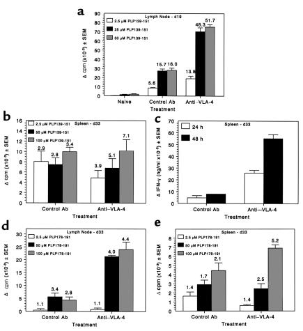
Figure 5.
Administration of anti–VLA-4 during ongoing R-EAE enhances proliferation and cytokine production to PLP139-151 and enhances epitope spreading to the relapse-associated PLP178-191 epitope. Spleen and lymph node cells from three mice per group were harvested at both 19 and 33 days after immunization from the experiment indicated in Figure 1c. (a) Viable lymph node cells (5 × 10⁵/well) from mice at day 19 after immunization (these mice had received three treatments with PS/2 beginning on day 14) were cultured with indicated concentrations of PLP139-151 for 4 days and proliferation was assessed by incorporation of 3H-TdR. 3H-TdR incorporation in cultures containing medium only were 1874 ± 286 and 1488 ± 334 for control and anti–VLA-4 groups, respectively. (b) Viable spleen cells (5 × 10⁵/well) from mice at day 33 after immunization (these mice had received nine treatments with PS/2 beginning on day 14) were cultured with indicated concentrations of PLP139-151 for 4 days and proliferation assessed by incorporation of 3H-TdR. 3H-TdR incorporation in cultures containing medium only was 4,199 ± 306 and 1,662 ± 248 for control and anti–VLA-4 groups, respectively. (c) Supernatants from the day 33 splenocyte cultures were harvested at 24 and 48 hours and analyzed for IFN-γ secretion by ELISA as described in Methods. Proliferative responses at day 33 after immunization to the relapse-associated PLP178-191 epitope were assessed from the lymph nodes (d) and the spleen (e). For d, 3H-TdR incorporation in cultures containing medium only was 2,214 ± 221 and 6,993 ± 1,274 for control and anti–VLA-4 groups, respectively. In e, medium-only 3H-TdR incorporation was 4,199 ± 306 and 1,662 ± 248 for control and anti–VLA-4 groups, respectively.

