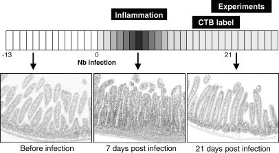
Figure 2. Experimental time course schematic illustrating the effect of Nb infection on jejunal histology.
Each image shows a typical haematoxylin and eosin-stained jejunal section at different time points throughout the experiment. Prior to exposure to Nb, the jejunum shows no sign of inflammation. One week post-Nb infection, there are clear signs of oedema and inflammatory cell infiltration in the jejunum. Three weeks post-Nb infection, the time point when experiments were performed, no oedema or inflammatory cell infiltrates are observed in the jejunum.

