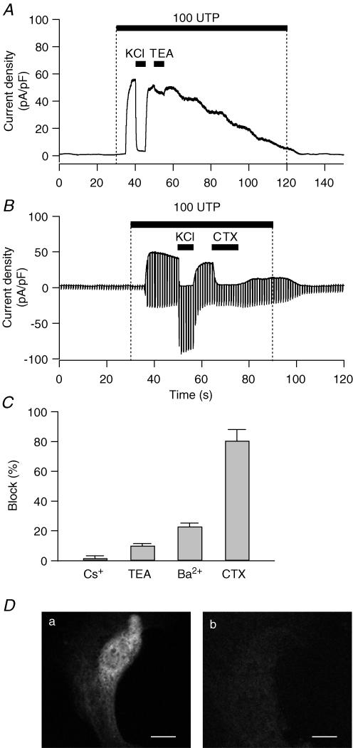
Figure 8. Pharmacology and immunostaining of the KCa channel in PDEC.
Cells were clamped at 0 mV in ruptured whole-cell configuration. A, effect of 10 mm TEA on K+ current activated by 100 μm UTP. To avoid non-specific effects caused by different osmolarity, the same amount of NaCl (10 mm) was added in the other solutions. B, effect of 100 nm CTX on K+ current mediated by 100 μm UTP. Membrane potential was held at 0 mV for 800 ms to measure KCa current and stepped to −80 mV for 200 ms to measure ClCa current every 1 s. C, effect of K+ channel blockers on PDEC KCa channels. Bar graph shows the inhibition observed with Cs+ (1 mm), TEA (10 mm), Ba2+ (5 mm), and CTX (100 nm). D, demonstration of IK1/SK4 channels in PDEC by immunofluorescence. Confocal images of cells treated with both primary and secondary antibody (a) or with secondary antibody alone (b). Scale bars indicate 10 μm.

