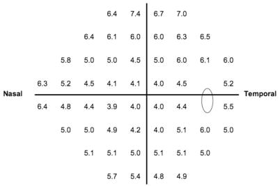
Figure 1.
Cutpoints (in decibels) at each of the 52 visual field locations used to identify an extreme reduction in sensitivity in the OHTS eyes corrected for age 45. Clinically, if the measured asymmetry for a patient at a particular point is larger than that presented at the same point in the figure, then the eye with the lower sensitivity should be flagged for increased risk of POAG.

