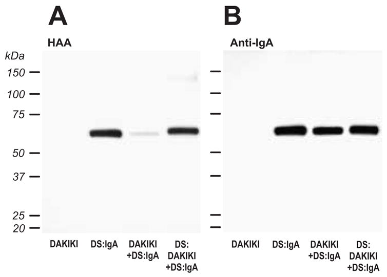
Figure 2. Sialylation of terminal GalNAc in the hinge region of IgA1 (Mce) by DAKIKI cell extracts.

Desialylated IgA1 (Mce) (DS:IgA) was incubated with a Golgi-enriched enzyme preparation from DAKIKI cells (DAKIKI+DS:IgA) and CMP-NeuAc and subsequently desialylated (DS:DAKIKI+DS:IgA). The samples, including DAKIKI Golgi preparations without added IgA1, were separated on SDS-PAGE, western-blotted onto a PVDF membrane and incubated with HAA lectin, that recognizes terminal GalNAc (A). To confirm that equivalent levels of IgA were loaded, the membrane was stripped and re-probed with an IgA-specific antibody (B).
