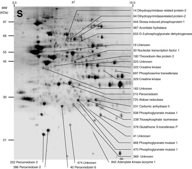
Figure 1. Representative image of a two-dimensional electrophoresis gel stained with Sypro Ruby, showing protein spots found in the soluble fraction (S) of normal rat medial vestibular nuclei (MVN) tissue.
Between 800 and 1000 spots were detected on individual gels using Phoretix 2005 two-dimensional gel analysis software (for details see Methods). Spot numbers were assigned by the software and synchronized between gels. Spots which showed significant changes in mean normalized volume after either sham surgery (compared to normal MVN) or unilateral labyrinthectomy (compared to sham-operated MVN) were selected for analysis, and are indicated. The protein content of the selected spots was determined by spot-picking, trypsin digestion and MALDI-TOF mass spectrometry of the tryptic peptides. Of 28 protein spots in the S fraction that showed significant changes, the identity of the protein content of 22 spots was established and is indicated, while the identity of the protein content of six spots could not be determined. The rectangular region D indicates the region of the gel illustrated in Fig. 3D.

