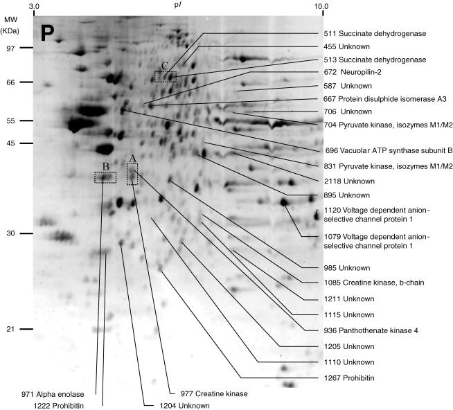
Figure 2. Representative image of a two-dimensional electrophoresis gel stained with Sypro Ruby, showing protein spots found in the pelleted fraction (P) of normal rat MVN tissue.
Due to the fractionation procedure used here, this fraction was expected to contain membrane-associated proteins. As in Fig. 1, protein spots which showed significant changes in mean normalized volume after either sham surgery (compared to normal MVN) or unilateral labyrinthectomy (compared to sham-operated MVN) were selected for analysis, and these are indicated. Of 26 protein spots in the P fraction that showed significant changes, the identity of the protein content of 15 spots was established and is indicated, while the identity of the protein content of 11 spots could not be determined. The rectangular regions A, B and C indicate the regions of the gel illustrated in Fig. 3A, B and C, respectively.

