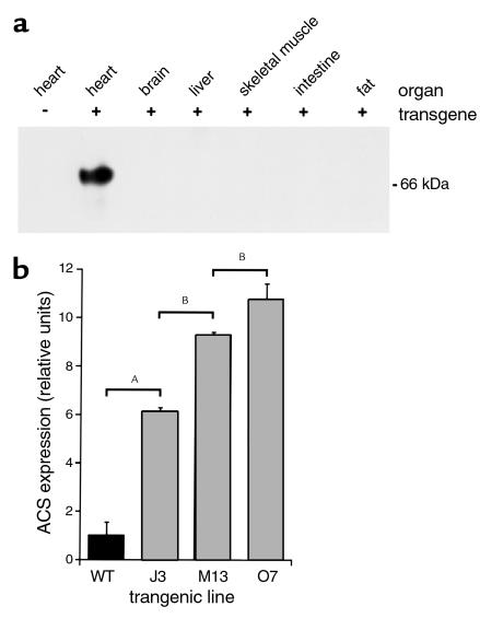
Figure 1.
Cardiac overexpression of murine ACS1 in MHC-ACS lines. (a) Membrane protein (20 μg) from various organs of an MHC-ACS O7 transgenic animal and nontransgenic littermate were separated by SDS-PAGE. Specific transgene expression was analyzed by Western blot, using a monoclonal anti-MYC Ab. (b) Membrane protein (20 μg) from hearts of 18-day-old MHC-ACS transgenic mice were analyzed by Western blot using rabbit polyclonal antisera directed against native murine ACS1 sequences. ACS1-specific signal was quantified and relative units of ACS1 expression is shown for wild-type (WT) and three independent transgenic lines (J3, M13, and O7). Data are reported as the mean ± SE. Differences among groups were compared by one-way ANOVA in conjunction with the post hoc Scheffé test (AP < 0.0001; BP < 0.01).

