Figure 7.
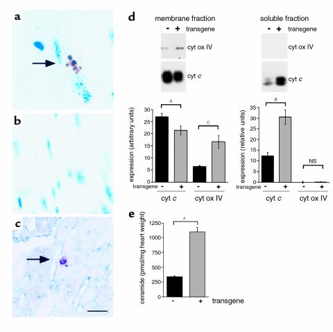
Evidence for lipoapoptosis in MHC-ACS hearts. (a–c) Cardiac ventricular tissues from a 18-day-old O7 transgenic mice (a and c) and from a nontransgenic control littermate (b) were fixed in formalin, embedded in paraffin, and sectioned. Tissues were stained for DNA fragmentation by a TUNEL protocol that stains apoptotic nuclei brown and allows visualization of myocyte striations. Double staining for α-sarcomeric actin was used to identify cardiomyocytes in c (myocyte cytoplasm blue). (d) Cardiac ventricular tissues from 21-day-old O7 transgenic mice and nontransgenic control littermates were flash-frozen, homogenized, and separated by differential density centrifugation to yield a membrane fraction (mitochondria) and soluble fraction (cytosol). Membrane protein (20 μg) and soluble protein (80 μg) were separated by SDS-PAGE and analyzed by Western blotting using an anti–cytochrome c (cyt c) Ab and an anti–cytochrome oxidase subunit IV (cyt ox IV) Ab. Bands were quantified using Molecular Analyst software, and relative units of expression are shown for wild-type (–) and transgenic (+) tissues. Data points are expressed as mean (minimum of four independent samples) ± SE. Nonpaired t test was used to compare groups (AP = 0.06, BP = 0.01, CP < 0.01). (e) Ceramide was measured in heart tissue from 18-day-old transgenic and wild-type animals using the diacylglycerol kinase assay and normalized for tissue weight. Data points are expressed as mean (minimum of five independent samples) ± SE. Statistical evaluation between groups was by Student’s t test (AP < 0.0005).

