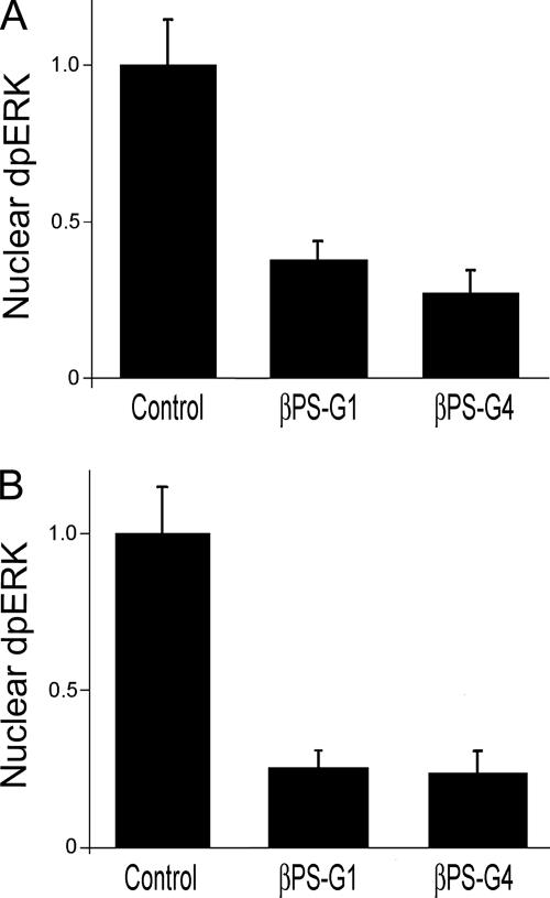
Figure 4.
Integrin mutations inhibit dpERK nuclear localization. Spreading (A) or insulin-stimulated (B) cells were stained for dpERK as described in Figure 2. Cells expressing mutations in integrin β subunits that alter cytoplasmic residues (G1) or ligand binding (G4) greatly reduce nuclear dpERK, even though these cells are spread to similar extents. (The βPS-G4 cells are spread independently of their integrins; see text.) Error bars indicate standard deviations from averages from three experiments; each experiment represents a minimum of 10 cells from seven different fields. Representative images of treated cells can be found in Supplemental Figure 3.

