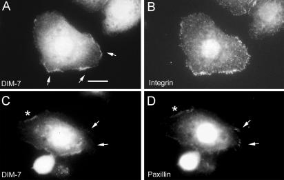
Figure 8.
DIM-7 and integrin colocalization. Drosophila S2 cells expressing αPS2βPS integrins were spread on a fragment of the ECM ligand Tiggrin, and fixed and stained to show the distributions of various components. (A and B) DIM-7 is found at high levels around the cell nucleus, and it also is localized at the cell periphery where concentrations of integrins (stained with anti-βPS) are mediating cell spreading (arrows). (C and D) DIM-7 is specifically absent from focal adhesions (visualized by high-density accumulations of paxillin, arrows) and the surrounding areas of the cell periphery. More diffuse paxillin and DIM-7 are seen along spreading edges of the cell (e.g., asterisks). Bar, 10 μm.

