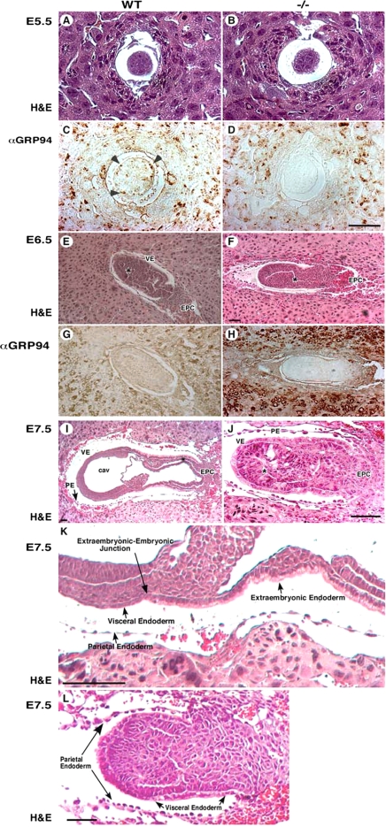
Figure 2.
Grp94−/− embryos fail to gastrulate. (A–H) Histological and immunostaining analysis of WT (left) and mutant (right) embryos. Transverse sections of E5.5 embryos (A–D), E6.5 embryos (E–H), or E7.5 embryos were stained either with hematoxylin and eosin (H&E, A,B and E,F) or mAb 9G10 (αGRP94, C,D and G,H). This antibody reacts with an epitope in the second, charged domain of GRP94. Arrowheads in C denote individual cells with high GRP94 expression. VE, visceral endoderm; EPC, ectoplacental cone. (E and F) Asterisk denotes the developing proamniotic cavity. (I–L) Histological analysis of E7.5 embryos. Transverse H&E-stained sections showing the lack of mesoderm formation and lack of cavitation in −/− embryos (J; denoted with * compared with WT embryos (I). PA, proamniotic cavity; EC, exoceolomic cavity; PE, parietal endoderm. The arrows in I–L mark the junctions between the embryonic visceral endoderm and the extraembryonic visceral endoderm. (K) Higher magnification image of the embryo in I, showing the cuboidal architecture of cells on the extraembryonic side of the endoderm junction and the squamous morphology of the endoderm cells on the embryonic side of the junction. (L) A similar view of a grp94−/− embryo, where the VE cells on both sides of the junction are cuboidal. Note also the lack of any evidence for proamniotic cavity. The PE cells do not look different in the mutant and WT embryos. All magnification bars, 50 μm.

