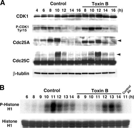
Figure 4.
Delayed activation of cyclin B/Cdk1 complex in toxin B–treated cells. (A) Immunoblot analysis for Cdk1, phosphoTyr15-Cdk1, Cdc25A, and Cdc25C. HeLa cells were collected every 2 h from 4 to 16 h after the second release with the toxin B addition at 8 h. Total cell lysates were prepared and subjected to SDS-PAGE and immunoblot analysis as indicated. Arrowheads indicate a phosphorylated form of each Cdc25 isoform. (B) Kinase assay of the cyclin B/Cdk1 complex. HeLa cells were collected at indicated times after the second release. Cell lysates were prepared and subjected to immunoprecipitation with antibody to cyclin B1. Immunoprecipitates were incubated with histone H1 in the presence of [γ-32P]ATP, and [32P]phosphorylated histone H1 was analyzed by SDS-PAGE and autoradiography.

