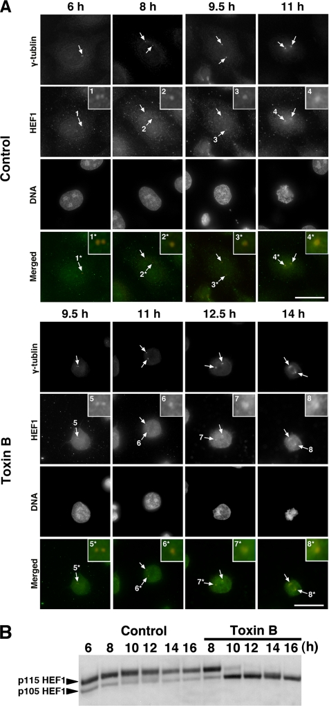
Figure 6.
Effects of toxin B treatment on localization and Ser/Thr phosphorylation of HEF1. (A) Immunofluorescence for HEF1. HeLa cells were fixed with methanol after pre-extraction with 0.5% Triton X-100 in PHEM buffer and stained for HEF1 (green), γ-tubulin (red), and DNA. Arrows indicate centrosomes, and insets show magnified views of the centrosomes indicated numbered arrows. Note that HEF1 was already present at the centrosome in the end of S phase (6 h) and increased its amount during G2 progression in control cells and that this increase was delayed in toxin B–treated cells. (B) Immunoblot for HEF1. HeLa cells were collected at indicated times after the release with toxin B addition at 8 h and were subjected to immunoblot for HEF1. Note that the toxin B treatment abolished p115 phosphorylated form of HEF1. Typical results of at least three independent experiments are shown. Bar, 10 μm.

